| MeSH term | MeSH ID | Detail |
|---|---|---|
| Goldenhar Syndrome | D006053 | 1 associated lipids |
| Lymphoma, Follicular | D008224 | 3 associated lipids |
| Supratentorial Neoplasms | D015173 | 1 associated lipids |
| Myopathy, Central Core | D020512 | 1 associated lipids |
| Rhabdomyosarcoma, Alveolar | D018232 | 2 associated lipids |
| Rhabdomyosarcoma, Embryonal | D018233 | 1 associated lipids |
| Rhabdoid Tumor | D018335 | 1 associated lipids |
| Rubinstein-Taybi Syndrome | D012415 | 1 associated lipids |
| Adenomyosis | D062788 | 1 associated lipids |
| Lymphoma, Large-Cell, Anaplastic | D017728 | 3 associated lipids |
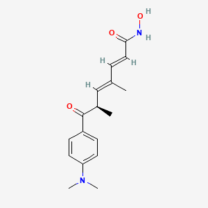
trichostatin A
Trichostatin is a lipid of Polyketides (PK) class. Trichostatin is associated with abnormalities such as Dentatorubral-Pallidoluysian Atrophy, PARAGANGLIOMAS 3, abnormal fragmented structure, Disintegration (morphologic abnormality) and Hyperostosis, Diffuse Idiopathic Skeletal. The involved functions are known as Acetylation, Cell Differentiation process, histone modification, Gene Silencing and Transcriptional Activation. Trichostatin often locates in CD41a, Hematopoietic System, Chromatin Structure, Blood and Endothelium. The associated genes with Trichostatin are SPI1 gene, CELL Gene, Chromatin, CXCR4 gene and DNMT1 gene. The related lipids are Butyrates, Promega, butyrate, Lipopolysaccharides and Steroids. The related experimental models are Knock-out, Mouse Model, Xenograft Model and Cancer Model.
Cross Reference
Introduction
To understand associated biological information of trichostatin A, we collected biological information of abnormalities, associated pathways, cellular/molecular locations, biological functions, related genes/proteins, lipids and common seen animal/experimental models with organized paragraphs from literatures.
What diseases are associated with trichostatin A?
trichostatin A is suspected in Infection, Morphologically altered structure, Ureteral obstruction, Photosensitization, Atherosclerosis, Hypertrophic Cardiomyopathy and other diseases in descending order of the highest number of associated sentences.
Related references are mostly published in these journals:
| Disease | Cross reference | Weighted score | Related literature |
|---|
Loading... please refresh the page if content is not showing up.
Possible diseases from mapped MeSH terms on references
We collected disease MeSH terms mapped to the references associated with trichostatin A
PubChem Associated disorders and diseases
What pathways are associated with trichostatin A
Lipid pathways are not clear in current pathway databases. We organized associated pathways with trichostatin A through full-text articles, including metabolic pathways or pathways of biological mechanisms.
Related references are published most in these journals:
| Pathway name | Related literatures |
|---|
Loading... please refresh the page if content is not showing up.
PubChem Biomolecular Interactions and Pathways
Link to PubChem Biomolecular Interactions and PathwaysWhat cellular locations are associated with trichostatin A?
Visualization in cellular structure
Associated locations are in red color. Not associated locations are in black.
Related references are published most in these journals:
| Location | Cross reference | Weighted score | Related literatures |
|---|
Loading... please refresh the page if content is not showing up.
What functions are associated with trichostatin A?
Related references are published most in these journals:
| Function | Cross reference | Weighted score | Related literatures |
|---|
What lipids are associated with trichostatin A?
Related references are published most in these journals:
| Lipid concept | Cross reference | Weighted score | Related literatures |
|---|
Loading... please refresh the page if content is not showing up.
What genes are associated with trichostatin A?
Related references are published most in these journals:
| Gene | Cross reference | Weighted score | Related literatures |
|---|
What common seen animal models are associated with trichostatin A?
Mouse Model
Mouse Model are used in the study 'Regulation of minichromosome maintenance gene family by microRNA-1296 and genistein in prostate cancer.' (Majid S et al., 2010), Mouse Model are used in the study 'Reversal of hypermethylation and reactivation of p16INK4a, RARbeta, and MGMT genes by genistein and other isoflavones from soy.' (Fang MZ et al., 2005) and Mouse Model are used in the study 'Histone deacetylase 3 mediates allergic skin inflammation by regulating expression of MCP1 protein.' (Kim Y et al., 2012).
Xenograft Model
Xenograft Model are used in the study 'Histone deacetylase inhibitors induce growth arrest and differentiation in uveal melanoma.' (Landreville S et al., 2012), Xenograft Model are used in the study 'Extended treatment with physiologic concentrations of dietary phytochemicals results in altered gene expression, reduced growth, and apoptosis of cancer cells.' (Moiseeva EP et al., 2007) and Xenograft Model are used in the study 'Retinoic acid and the histone deacetylase inhibitor trichostatin a inhibit the proliferation of human renal cell carcinoma in a xenograft tumor model.' (Touma SE et al., 2005).
Cancer Model
Cancer Model are used in the study 'Plasma pharmacokinetics and metabolism of the histone deacetylase inhibitor trichostatin a after intraperitoneal administration to mice.' (Sanderson L et al., 2004).
Related references are published most in these journals:
| Model | Cross reference | Weighted score | Related literatures |
|---|
Loading... please refresh the page if content is not showing up.
NCBI Entrez Crosslinks
All references with trichostatin A
Download all related citations| Authors | Title | Published | Journal | PubMed Link |
|---|---|---|---|---|
| Guo W et al. | Abrogation of TGF-beta1-induced fibroblast-myofibroblast differentiation by histone deacetylase inhibition. | 2009 | Am. J. Physiol. Lung Cell Mol. Physiol. | pmid:19700647 |
| Popova AP et al. | Autocrine production of TGF-beta1 promotes myofibroblastic differentiation of neonatal lung mesenchymal stem cells. | 2010 | Am. J. Physiol. Lung Cell Mol. Physiol. | pmid:20190033 |
| Alamdari N et al. | Sepsis and glucocorticoids upregulate p300 and downregulate HDAC6 expression and activity in skeletal muscle. | 2010 | Am. J. Physiol. Regul. Integr. Comp. Physiol. | pmid:20538901 |
| Yang H et al. | Dexamethasone-induced protein degradation in cultured myotubes is p300/HAT dependent. | 2007 | Am. J. Physiol. Regul. Integr. Comp. Physiol. | pmid:16973938 |
| Kroening S et al. | Characterization of connective tissue growth factor expression in primary cultures of human tubular epithelial cells: modulation by hypoxia. | 2010 | Am. J. Physiol. Renal Physiol. | pmid:20032117 |
| Yu Z and Kone BC | Targeted histone H4 acetylation via phosphoinositide 3-kinase- and p70s6-kinase-dependent pathways inhibits iNOS induction in mesangial cells. | 2006 | Am. J. Physiol. Renal Physiol. | pmid:16174862 |
| Noh H et al. | Histone deacetylase-2 is a key regulator of diabetes- and transforming growth factor-beta1-induced renal injury. | 2009 | Am. J. Physiol. Renal Physiol. | pmid:19553350 |
| Pang M et al. | Inhibition of histone deacetylase activity attenuates renal fibroblast activation and interstitial fibrosis in obstructive nephropathy. | 2009 | Am. J. Physiol. Renal Physiol. | pmid:19640900 |
| Marumo T et al. | Histone deacetylase modulates the proinflammatory and -fibrotic changes in tubulointerstitial injury. | 2010 | Am. J. Physiol. Renal Physiol. | pmid:19906951 |
| Dong G et al. | Inhibitors of histone deacetylases suppress cisplatin-induced p53 activation and apoptosis in renal tubular cells. | 2010 | Am. J. Physiol. Renal Physiol. | pmid:19889954 |